
High-end image documentation system for eye diagnostics using digital slit lamp photography
A deep look into the eye
The slit lamp examination is one of the most important diagnostic techniques in ophthalmology. It enables a detailed examination of the anterior, middle and posterior segment of the eye. Ophthalmologists can use it to detect the smallest changes, anomalies or damage. This procedure is used for the early detection and monitoring of the progression of eye diseases such as corneal injuries, eye infections, retinal detachment or macular degeneration. However, the eye, with its rapid movements, is a challenging subject to photograph; motion blur and shaking are typical image errors. Fast and reliable slit lamp documentation systems are needed to make diagnostics easier for ophthalmologists and opticians, improve the workflow and at the same time shorten examination times. They must provide meaningful images and be user-friendly and ergonomically designed.

The German company OCULUS Optikgeräte GmbH develops instruments for eye diagnostics for ophthalmologists, optometrists and opticians. Part of the extensive portfolio: one of the world's smallest and lightest image documentation systems for slit lamps. A powerful, high-resolution USB3 Vision industrial camera from IDS is integrated - especially suitable for applications in medical technology and microscopy.

"Only a fast camera delivers low-noise images in difficult shooting situations at the eye"
Efficient system
The OCULUS ImageCam®3 universal slit lamp image documentation system is not only intuitive and easy to use, but also sets high standards in digital slit lamp photography. This includes an outstanding field of view that enables highly precise diagnoses of the anterior, middle and posterior segments of the eye. The images are captured by a particularly low-light, high-performance IDS camera from the USB3 uEye+ CP family. "Only a fast camera delivers low-noise images in the difficult recording situations at the eye," explains Michael Moos, Product Manager at OCULUS, the selection of the camera model.

Another important requirement criterion is speed. "The advanced camera features enable continuous shooting at up to 60 frames per second. Among other things, these series recordings make it possible to record the eye during breaks in movement. The innovative frame-out-of-video function enables simple documentation of the entire examination process at the slit lamp, whereby the best quality individual images can then be selected for evaluation," says Michael Moos. The OCULUS ImageCam® 3 makes this possible without any loss of quality and in a minimum of time.
Powerful camera
The IDS camera from the CP family is predestined for use in medical technology, as it offers extensive pixel pre-processing and has an internal 120 MB image memory for buffering image sequences. This enables a high data rate of 420 MByte/s, low CPU load and easy integration. The Sony Pregius IMX265 in the model used here is considered one of the best CMOS image sensors in the 3 MP class. The USB3 Vision industrial camera U3-3270CP Rev.2.2 with the 1/1.8" global shutter sensor thus achieves a resolution of 3.19 megapixels (2064 x 1544 px). "In this case, however, the image is scaled down using AOI to achieve a significantly higher frame rate," explains Phillip Schissler, Sales Manager Medical and Microscopy at IDS. OCULUS has integrated the camera with the IDS peak software development kit. "IDS peak allows users to test camera functions in detail and optimize them for your own applications," says the IDS Medical expert.
However, the camera is not only recommended for medical technology and microscopy in terms of sensitivity, dynamic range and linearity. In addition to the required light intensity and speed, the size of the camera was also a decisive selection criterion for the model. At around 50 grams, the camera's small 29 x 29 x 29 millimeter magnesium housing is as light as it is robust, underlining its suitability for space-critical applications.

Facilitated slit lamp diagnostics
In order to deliver optimum diagnostic images, the system includes a high-quality beam splitter in addition to the camera. A beam splitter divides the light between the camera and the eyepiece of the slit lamp to simultaneously illuminate and view the eye, allowing a detailed examination of each eye segment. The beam splitter of the OCULUS system has a purely mechanical iris diaphragm that significantly increases the depth of field, regardless of the position of the pathological findings. It can also be adapted to all commercially available slit lamps. "The camera unit and the beam splitter are extremely small and light. This means in daily practice: It is barely noticeable, is very easy to attach and delivers images like no other in this size. This makes daily slit lamp diagnostics easier in the truest sense of the word," says the manufacturer.

In terms of image quality, reliability and long-term availability, IDS stands for the highest quality standards in laboratory and diagnostics.
The slit lamp photos of the OCULUS ImageCam® 3 enable objective documentation of eye conditions in order to monitor the progress of diseases and compare treatments. Patients also benefit from the image documentation system. The visual references of the diagnosis created in this way help them to better understand their condition and the doctors' treatment plan. The medical findings can be saved and archived accordingly.
Outlook
Innovative image processing systems such as OCULUS ImageCam®, which use powerful industrial cameras to deliver informative, high-contrast images with high depth of field, help to improve diagnostic accuracy, efficiency and patient care in ophthalmology. Artificial intelligence (AI) is increasingly being integrated into the analysis of slit lamp images to automatically detect diseases, support diagnostic decisions, improve the workflow for doctors and develop new treatment methods.
OCULUS Optikgeräte GmbH
The company OCULUS Optikgeräte GmbH has been a partner for ophthalmologists, optometrists and opticians worldwide for 125 years. The company develops first-class instruments for eye diagnostics for this clearly defined group of users. The measure of all things is the high demands of customers and the continuous further development of technologies. Over 55% of OCULUS' sales are realised in foreign markets. More than 400 employees work at the OCULUS headquarters. More than 60 % of them work in research and development, production and customer service. With 12 company-owned subsidiaries and more than 200 wholesalers in over 80 countries, OCULUS is at its customers' service around the globe. Founded in 1895, the family business is now jointly managed by the third and fourth generations.
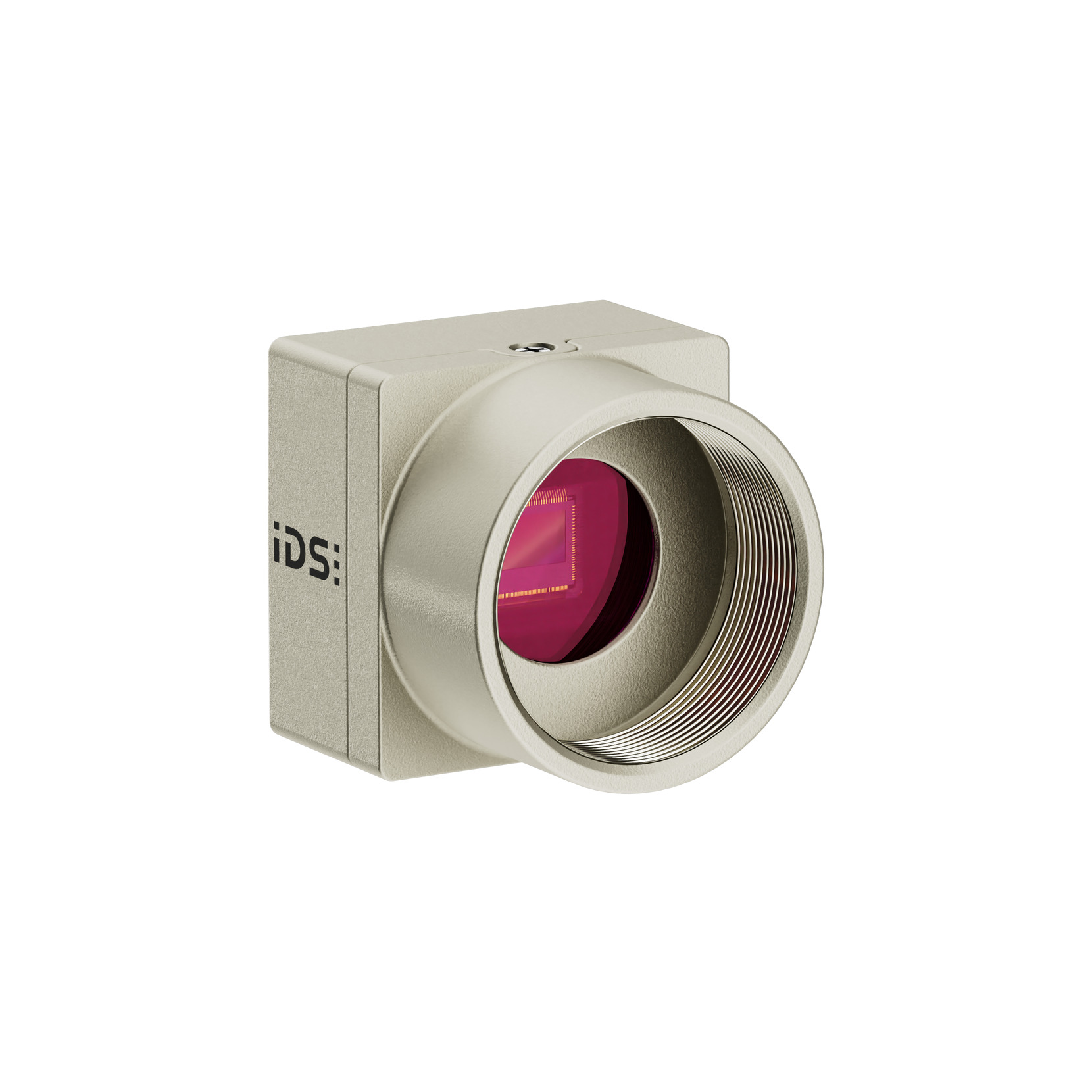
uEye CP
Model used: U3-3270CP Rev.2.2

For over ten years, she has been creating press releases and application reports and designing corporate topics and technical product communication with corresponding expertise. Thanks to her experience in strategic B2B communication, she formulates precise messages and delivers well-founded, technically robust content - always with a focus on authenticity and clarity.
Your project
How can we support you in your project? Together we will find the right solution for you!
Vision Channel
Videos and live sessions about machine vision.
Newsletter
Stay up to date and subscribe to our newsletter.
Applications
Discover how industrial cameras are shaping the future.